A working reference for the Technical Course: landmark datasets with specifications and access routes, the software landscape by workflow stage, benchmarks, and the curation schema for adding entries.
How to use this page
This is a lookup table, not a lesson. Come here when you need to answer one of:
Which public dataset can answer my question? → §1
What tool do I need at this pipeline stage? → §2
How do I evaluate a segmentation method? → §3
How do I add something to this atlas? → §4
Specifications below are as published and are approximate where releases have been
revised. Always confirm current figures and access terms against the primary source
before citing them — dataset sizes in particular change as proofreading continues
(Unit 04 §2).
1. Landmark datasets
Invertebrate
Dataset
Scale
Modality / resolution
What it is good for
Access
C. elegans (White et al. 1986; Cook et al. 2019; Witvliet et al. 2021)
302 neurons; whole animal; a developmental series across maturation
ssTEM
The complete-nervous-system reference case. Witvliet’s series is the best available data on how connectivity changes with development
WormWiring, WormAtlas
Larval Drosophila brain (Winding et al. 2023)
~3,000 neurons; ~550,000 synapses; whole brain
ssTEM
A whole brain small enough for exhaustive graph analysis; bilateral matching studies
CATMAID instances; published supplements
Drosophila hemibrain (Scheffer et al. 2020)
~25,000 neurons; ~20 million synapses; central brain
FIB-SEM, near-isotropic 8 nm
Cell-type census; the cleanest large connectome for analysis teaching, because isotropy makes tracing quality high
neuPrint
FAFB / FlyWire (Zheng et al. 2018; Dorkenwald et al. 2024)
Whole adult brain; ~139,000 neurons; ~54.5 million synapses
ssTEM, 4 × 4 × 40 nm
The first whole-brain connectome of a behaviorally complex animal; community-proofread
FlyWire (registration required); CAVE
Drosophila male adult nerve cord (MANC)
~23,000 neurons
FIB-SEM
Motor circuits; connecting brain to periphery
neuPrint
Vertebrate
Dataset
Scale
Modality / resolution
What it is good for
Access
Mouse retina (e2198 and relatives) (Briggman, Helmstaedter, Denk)
~10⁵–10⁶ µm³
SBEM
Structure-function in a well-characterized circuit; direction selectivity
Published; some via community portals
Kasthuri saturated reconstruction (Kasthuri et al. 2015)
~1,500 µm³ mouse neocortex, densely reconstructed
ssSEM (ATUM)
The reference for what dense, saturated reconstruction means and costs
Open Connectome / BossDB
Hippocampal CA1 resource (Harris et al. 2015)
Dense neuropil volume
ssTEM
Spine and synapse ultrastructure; a standard teaching set for Units 05–06
Published resource
MICrONS (Allen Institute, Baylor, Princeton; 2025 release)
~1 mm³ mouse visual cortex; ~200,000 cells; ~500 million synapses
The reference functional-connectomics dataset. The co-registration is what makes it unique
CAVE / caveclient; MicronsBinder notebooks
H01 human cortex (Shapson-Coe et al. 2024)
~1 mm³ human temporal cortex; ~57,000 cells; ~150 million synapses; ~1.4 PB
ssTEM, ~4 × 4 × 30 nm
Human tissue at synapse resolution; species comparison
Google/Lichtman lab public release; Neuroglancer
Larval zebrafish whole brain (Hildebrand et al. 2017)
Whole brain, larval
ssEM
Whole-vertebrate-brain scale in a tractable organism
Published resource
MouseConnects / HI-MC (NIH BRAIN CONNECTS)
Scaling toward whole mouse brain
Volume EM
The current flagship scaling effort; see the case study
Program resources
Choosing among them
If your question is about…
Start with
Whole-brain graph structure in a behaving animal
FlyWire, or larval Drosophila for exhaustive analysis
Cell types and their connectivity, with clean tracing
Hemibrain
Structure–function relationships
MICrONS
Human-specific features
H01
Development of connectivity
C. elegans developmental series
What dense reconstruction actually requires
Kasthuri 2015
Teaching ultrastructure reading
Harris CA1 resource; any of the above in Neuroglancer
Before proposing new acquisition, check this table. A large fraction of good
connectomics questions can be answered by re-analysis of existing public data. The
cost difference is not marginal: acquiring a new mm-scale volume is a multi-year,
multi-million-dollar program, while re-analyzing one is a compute bill and your
time. Work the comparison out for your own question with Unit 03’s cost arithmetic
before assuming you need new data. See Unit 02’s common errors.
2. Software, by workflow stage
Stage
Tool
What it does
Viewing
Neuroglancer
The standard browser-based viewer for petascale volumes, meshes, and annotations
webKnossos
Viewing, annotation, and proofreading with collaborative features
Data access
CloudVolume
Python access to precomputed/chunked volumes
caveclient
Client for CAVE — segmentation, synapse tables, materialization versions
Declarative subgraph/motif queries over connectomes
Hosting
BossDB
Community archive for volumetric neuroscience data
3. Benchmarks and evaluation
Benchmark
What it evaluates
Notes
SNEMI3D
3D neurite segmentation
Long-standing reference challenge; small volume
CREMI
Neuron segmentation and synaptic partner identification in Drosophila EM
Includes synaptic partner assignment, which most benchmarks omit
ISBI 2012
2D membrane segmentation
Historical; useful for teaching, not representative of current difficulty
Caution when reading benchmark results. Scores on small, well-prepared benchmark
volumes systematically overstate performance on production data, which contains
artifacts (Unit 03), rare morphologies, and volume boundaries that benchmarks exclude.
When evaluating a method for your project, the question is not its leaderboard
position but its error rate on your tissue — which means running it on a
representative sub-volume of your own data (Unit 03 §3, the pilot reconstruction rule).
Report metrics as described in Unit 08 §3: at minimum VI decomposed into split and
merge components, plus one tracing-oriented metric such as ERL, plus the effect on
your endpoint.
known_limits — one concise sentence on the technical boundary
mapped_units — technical-track unit slugs this supports
The known_limits field is the one most often left blank and the one most worth
filling. An atlas of tools without limits is advertising.
Curation policy
Add only resources with a clear technical contribution or benchmark value.
Mark superseded methods as historical rather than deleting them, when they remain
pedagogically useful — the history of a method often explains its assumptions.
Prefer resources with reproducible artifacts: data, code, or an explicit protocol.
Re-review on schedule; retire stale links.
Every entry links to at least one unit, so the atlas stays connected to teaching
rather than becoming an orphaned bibliography.
Quality-control checks
Link health check on every external URL
Metadata completeness against the required schema
Duplicate detection (the same method published across multiple venues)
Coverage audit, so no single workflow stage dominates
Visual context set
This page is a lookup table and this panel is deck context, not reference material — nothing here should be cited. Use it instead to rehearse the habit §1 asks for: confirm every specification against the primary source, because dataset sizes and cell counts change as proofreading continues.
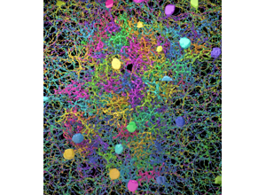
Module14 L3 S03-02: The opening of the source deck’s reference stream. Treat any figure it carries as provisional — volume sizes, cell counts, and synapse counts in this field are release-dependent, so check them against the primary source before citing (Unit 04 §2).
Module14 L3 S10: Mid-stream references. Use it to test the curation standard in §4: for each resource named, ask whether you could write its known_limits sentence. That field is the one most often left blank, and an atlas of tools without limits is advertising.
Module14 L3 S19: Closing references. Check them against the choosing table in §1 before proposing new acquisition — a large share of good connectomics questions can be answered by re-analysis of existing public data, at a small fraction of the cost.
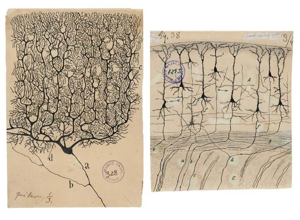
Techtalk S44: Developmental motif comparison, cross-referenced here from Unit 09. Use it as the pointer into the C. elegans developmental series in §1 — the dataset to reach for when the question is how connectivity changes with maturation rather than what it is in one adult.
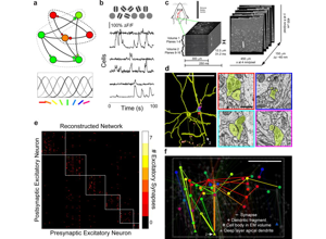
Module14 L3 S13: Functional recordings, an EM volume, reconstructed neurons and a connectivity matrix — several views of one dataset in one figure. Fill in the §4 modality and effective_resolution fields for it and the problem shows: an entry has to say which view it indexes, because the calcium data and the EM data have different resolutions and support different claims.
Module14 L3 S08: A circuit figure from the fly visual system, with reconstructed neurons above and a wiring diagram naming cell types L1, Mi1 and Tm3. Try to write its entry from the figure alone and you cannot: the citation and dataset_or_code_access fields need the paper, not the slide. That gap is why this panel is context and §1 is the reference.
Module14 L3 S05: A region-level brain network: named cortical areas as nodes, drawn from above and from the side. None of the datasets in §1 works at this scale. It marks the atlas's boundary — synapse-resolution volumes — and the macroscale methods that produce figures like this are placed on Unit 02's modality chart instead.
Attribution: module14 lesson3 and neuroAI source decks (historical/context visuals).
Mini-lab: curate one entry (30 minutes)
Add one resource to the atlas. Produce:
All required metadata fields, complete.
One sentence on the technical contribution — what can you now do that you could not?
One sentence on the limitation or failure context — when does this not work?
The unit or units it supports, with a sentence on how.
Rubric.Proficient: all fields present and accurate. Strong: the limitation
sentence is specific enough that a reader could predict a failure case from it, and
the unit mapping identifies where in that unit’s workflow the resource fits.
References
§4 of this page requires every contributed entry to carry “a standardized
citation string, with DOI”. This section is that rule applied to the page
itself. Every dataset named in §1 is here, with the published reference
rather than its preprint — several of these circulated as bioRxiv preprints for
years and are still cited that way.
Invertebrate
White JG, Southgate E, Thomson JN, Brenner S (1986). The structure of the nervous system of the nematode Caenorhabditis elegans. Philosophical Transactions of the Royal Society B 314:1-340. 10.1098/rstb.1986.0056
Cook SJ, Jarrell TA, Brittin CA, et al. (2019). Whole-animal connectomes of both Caenorhabditis elegans sexes. Nature 571:63-71. 10.1038/s41586-019-1352-7
Witvliet D, Mulcahy B, Mitchell JK, et al. (2021). Connectomes across development reveal principles of brain maturation. Nature 596:257-261. 10.1038/s41586-021-03778-8
Winding M, Pedigo BD, Barnes CL, et al. (2023). The connectome of an insect brain. Science 379:eadd9330. 10.1126/science.add9330
Scheffer LK, Xu CS, Januszewski M, et al. (2020). A connectome and analysis of the adult Drosophila central brain. eLife 9:e57443. 10.7554/eLife.57443
Zheng Z, Lauritzen JS, Perlman E, et al. (2018). A complete electron microscopy volume of the brain of adult Drosophila melanogaster. Cell 174:730-743. 10.1016/j.cell.2018.06.019
Dorkenwald S, Matsliah A, Sterling AR, et al. (2024). Neuronal wiring diagram of an adult brain. Nature 634:124-138. 10.1038/s41586-024-07558-y
Takemura S, Hayworth KJ, Huang GB, et al. (2024). A connectome of the male Drosophila ventral nerve cord. eLife 13:RP97769. 10.7554/eLife.97769
Vertebrate
Briggman KL, Helmstaedter M, Denk W (2011). Wiring specificity in the direction-selectivity circuit of the retina. Nature 471:183-188. 10.1038/nature09818
Helmstaedter M, Briggman KL, Turaga SC, et al. (2013). Connectomic reconstruction of the inner plexiform layer in the mouse retina. Nature 500:168-174. 10.1038/nature12346
Kasthuri N, Hayworth KJ, Berger DR, et al. (2015). Saturated reconstruction of a volume of neocortex. Cell 162:648-661. 10.1016/j.cell.2015.06.054
Harris KM, Spacek J, Bell ME, et al. (2015). A resource from 3D electron microscopy of hippocampal neuropil from adult rat stratum radiatum. Scientific Data 2:150046. 10.1038/sdata.2015.46
Hildebrand DGC, Cicconet M, Torres RM, et al. (2017). Whole-brain serial-section electron microscopy in larval zebrafish. Nature 545:345-349. 10.1038/nature22356
The MICrONS Consortium (2025). Functional connectomics spanning multiple areas of mouse visual cortex. Nature 640:435-447. 10.1038/s41586-025-08790-w
Shapson-Coe A, Januszewski M, Berger DR, et al. (2024). A petavoxel fragment of human cerebral cortex reconstructed at nanoscale resolution. Science 384:eadk4858. 10.1126/science.adk4858
MouseConnects / HI-MC is an in-progress BRAIN CONNECTS program rather than a
released dataset; see the case study
for its current state. Tool entries in §2 link to their own documentation, which
is the citable source for a version.
Curate an entry with complete metadata and explicit limitations.
Assign evidence maturity and review cadence for each resource.
Capability development brief
Capability target: Curate a trustworthy connectomics reference atlas with transparent metadata and evidence quality signals.
Required expertise
Information scientist (taxonomy and metadata schemas)
Domain editor (method and dataset quality review)
Reproducibility lead (citation and versioning standards)
Core concepts to teach
Metadata completeness: Minimum descriptive fields required for responsible reuse.
Evidence grading: Explicit quality and maturity signals for methods, datasets, and tools.
Reference maintenance: Scheduled review and change tracking to keep entries current.
Capability exercise
Atlas Entry Sprint - Produce publishable reference entries with complete metadata and quality notes. The mini-lab above is the graded version of this exercise; do that one.
Assessment artifacts
Atlas entry template and quality checklist.
Curated starter index with versioned citations.
Related concepts
Reproducibility and Reference Curation
Curate methods, datasets, and tools with metadata completeness and explicit limitations.