How to actually read an EM image: the organelle catalog with sizes, the minimum criteria for calling a synapse, Gray type I vs II, and a calibrated confidence protocol.
Key community resources for this unit:
Peters, Palay & Webster, The Fine Structure of the Nervous System
The standard reference atlas/textbook for identifying neuronal organelles and ultrastructure in EM (3rd ed., Oxford University Press, 1991).
Self-study ~3.75 h: about 2.5 h of reading plus the 75 min studio activity. Taught: a 95 min session, per the lecture plan. Deck: the unit’s slide deck is scoped to 60 min and does not follow the plan slide for slide.
Prerequisites
Units 01–03. Unit 03’s artifact catalog in particular — you cannot distinguish biology from artifact without it.
You need
A public EM volume open in Neuroglancer
You finish with
Calibrated compartment and synapse calls with justified confidence tiers, plus a personal cue-reliability ranking
Everything downstream depends on someone being able to look at a patch of grayscale
noise and say correctly what it is. Segmentation networks are trained on those
judgments. Proofreading decisions rest on them. Every synapse count in every
connectomics paper traces back to a human who decided that a particular smudge was a
postsynaptic density.
This unit teaches the actual visual cues, with sizes. Not “use organelle evidence” —
which organelles, how big, and what they rule out.
What you’ll be able to do
Name the major organelles visible in EM, with approximate sizes, and say which compartment each implies.
Apply the three minimum criteria for calling a chemical synapse, and refuse to call one when a criterion is missing.
Distinguish Gray type I from type II morphology and state the inference each licenses — and its limits.
Assign a calibrated confidence tier with a stated evidence chain.
Diagnose your own errors by cue, not just by count.
1. The organelle catalog
This is the reference table. Sizes are approximate and vary with preparation, but the
relative sizes and the presence/absence patterns are what you actually use.
Structure
Size
Appearance in EM
Found in
Practically absent from
Synaptic vesicle, clear round
35–50 nm
Small circular profiles, clear lumen, clustered
Presynaptic terminals
Dendrites, glia
Synaptic vesicle, pleomorphic/flattened
~35–50 nm
Oval or flattened profiles; shape is partly a fixation artifact but is diagnostically useful
Inhibitory terminals
Excitatory terminals
Dense-core vesicle
80–120 nm
Circular with a dark core
Peptidergic/monoaminergic terminals; also in transit along axons
—
Postsynaptic density (PSD)
30–50 nm thick, 200–800 nm wide
Dark, granular thickening under the postsynaptic membrane
Postsynaptic side
Presynaptic side
Synaptic cleft
20–30 nm (asymmetric); ~15–20 nm (symmetric)
Uniform-width gap with parallel membranes, often with faint cross-bridges
Between synaptic partners
Random appositions have variable-width gaps
Microtubule
~25 nm outer diameter
Tubule in longitudinal section; small ring in cross-section
Dendrites (abundant, in loose parallel arrays); axons (present, more regularly spaced)
Mature spine heads; most glial processes
Neurofilament
~10 nm
Fine filaments, often in bundles
Axons, especially myelinated
Spines
Mitochondrion
0.2–1 µm diameter, variable length
Double membrane with cristae
Everywhere except thin spine necks and the thinnest processes
—
Rough ER / polyribosomes
Ribosome ~25 nm
Studded membrane sheets; ribosome rosettes
Soma, proximal dendrites, dendritic shafts
Axons — a workhorse discriminator
Golgi apparatus
~1 µm stack
Stacked flattened cisternae with vesicles
Soma, proximal dendrite
Axons
Smooth ER / spine apparatus
Laminae ~30 nm
Tubules; in spines, stacked laminae with dense material between
Dendrites; spine apparatus in a minority of (mostly large) spines
—
Multivesicular body
200–500 nm
Membrane-bound body containing small internal vesicles
Everywhere; enriched in dendrites
—
Glycogen granule
20–30 nm
Very dark small particles, clustered
Astrocytes — near-diagnostic
Neurons
Myelin
10–20 lamellae
Regular concentric dark lamellae
Around myelinated axons
—
AIS undercoating
~20 nm dense layer
Granular density beneath the axolemma, plus fasciculated microtubules
Axon initial segment (~20–60 µm from soma), nodes of Ranvier
Everywhere else
The two highest-value entries for a beginner are the ones in bold logic:
ribosomes rule out axon, and glycogen granules indicate astrocyte. Those two facts
alone resolve a large share of early-annotator confusion.
Check yourself
A process ~400 nm across contains a mitochondrion, several microtubules, and
what look like a few ribosome rosettes. No vesicles, no PSD visible in this plane.
Best call, and what would raise your confidence?
Probable dendrite (or a proximal dendritic branch), medium confidence. The
ribosome rosettes are the strongest single cue — axons are effectively free of
polyribosomes in standard EM connectomics practice, so their presence argues
strongly against axon. The microtubules and mitochondrion are consistent but not
discriminating; both compartments have them.
To raise confidence, look for cues that are independent of the ribosome call:
Scroll through z and look for spines emerging from the process. A spine with a
head and neck is close to definitive for dendrite.
Look for incoming asymmetric synapses where this process is postsynaptic — a
PSD on this process means it is receiving, which is dendritic (or somatic).
Follow the process toward larger caliber and check whether it thickens toward a
soma. Dendrites taper with distance from soma; axons maintain caliber.
Note the reasoning pattern, which is the transferable skill: do not stack more
of the same kind of evidence. Three microtubule observations are one piece of
evidence. A ribosome plus a spine plus a taper is three.
2. Calling a synapse: the three criteria
A chemical synapse in EM requires all three:
A presynaptic vesicle cluster — a group of vesicles gathered at the membrane
facing the partner. Not scattered vesicles somewhere in the profile; clustered at
the apposition.
A synaptic cleft — parallel membranes with a consistent gap, wider than the
~10–20 nm typical of casual membrane apposition, and of uniform width across the
contact.
A postsynaptic density — a visible dark thickening on the receiving side.
And a fourth practical requirement that experienced annotators treat as
non-negotiable:
Persistence across sections. The features should be visible on more than one
consecutive section. A single-section “synapse” at 40 nm z-resolution is one
sample of a structure that is typically 200–500 nm wide — if it is real, you should
see it two to five times.
The single most common beginner error is calling a synapse from dark contrast
alone. Dark contrast at a membrane can be: a genuine PSD, a tangentially cut
membrane (very common — a membrane sliced obliquely looks thick and dark), staining
precipitate, a glial apposition, or a puncta adherens / adherens junction. Criterion
1 is what separates these: no vesicles, no synapse.
Adherens junctions: the classic false positive
Puncta adherentia have symmetric densities on both sides and no vesicle cluster.
They look convincing at first glance. The tell is symmetry plus the absence of a
vesicle pool.
Gray type I vs type II
Type I (asymmetric)
Type II (symmetric)
PSD
Thick, prominent, clearly asymmetric
Thin, roughly equal to the presynaptic density
Cleft
Wider, ~20–30 nm
Narrower, ~15–20 nm
Vesicles
Round, clear
Pleomorphic / flattened
Usual location
Dendritic spines; some shafts
Shafts, soma, AIS
Usual inference
Excitatory (glutamatergic)
Inhibitory (GABAergic)
The inference is a Bin B claim in the Unit 01 sense. It is a well-supported
statistical association, not an identity. Known complications: vesicle shape depends
on fixation and can be unreliable; some glutamatergic synapses onto interneuron shafts
appear less asymmetric; neuromodulatory terminals do not fit the dichotomy at all.
Therefore: write “putatively excitatory (asymmetric)” rather than “excitatory”,
and where the claim matters, corroborate with the identity of the presynaptic cell
type, which is usually the stronger evidence.
Check yourself
You see a dark thickening between two processes. On the section above and
below, the thickening is absent. One process contains a mitochondrion; neither shows a
clear vesicle cluster. Call?
Not a synapse — do not annotate one. Criterion 1 fails (no vesicle cluster) and
criterion 4 fails (not persistent).
The most likely explanations are a tangentially sectioned membrane or a
non-synaptic apposition. The presence of a mitochondrion is not evidence either
way; mitochondria are everywhere.
The correct output is a negative call, and if the region is ambiguous enough to
have cost you time, log it as an uncertain patch so it enters the calibration set.
Negative calls are data. An annotator who never says “no” is not calibrated.
A bouton contains round clear vesicles, makes a contact with a thick PSD onto
a spine head, and *also* contacts a nearby dendritic shaft with a thin symmetric
density. What is going on?
Most likely the second contact is not a synapse from this bouton — check for a
vesicle cluster at that apposition specifically. A single terminal’s vesicle pool
can sit near several membranes; only the apposition with an adjacent vesicle
cluster and a cleft counts.
If a vesicle cluster genuinely is present at both, you have a multi-synaptic
bouton, which is real and common. Note that one terminal making both an
asymmetric contact onto a spine and a symmetric contact onto a shaft would be
unusual and worth flagging for expert review — it may indicate a merge error
that has fused two different axons into one object. This is a good example of
ultrastructural reading catching a segmentation error: the biology looks wrong, so
suspect the segmentation.
3. Compartment cues: a decision protocol
Work in this order. The order matters — cheap, reliable cues first.
Step 1 — Local geometry. Diameter and its variation. Boutons are swellings
connected by thin intervaricose segments; dendritic shafts have relatively smooth
caliber; spine heads sit on necks.
Step 2 — Organelles present, and just as important, organelles absent.
Ribosomes present → not axon. Glycogen granules → astrocyte. Vesicle cluster →
presynaptic. Absence is evidence when the structure would be visible if present at
this magnification and plane.
Step 3 — Synaptic role. Does the process bear PSDs (receiving) or vesicle clusters
(sending)? Many processes do both, but the balance is informative.
Step 4 — Continuity across sections. This is where most single-plane calls get
overturned. Scroll. A process that looked like a bouton may be a dendritic varicosity;
a “vesicle cluster” may be a tangential slice through something else.
Step 5 — Neighborhood. What is around it? An axon in a myelinated bundle, a
process wrapping a capillary, a profile inside a glial sheath — context frequently
settles calls that local features cannot.
Step 6 — Assign confidence with a stated evidence chain.
The confidence tiers, defined operationally
Tiers are useless unless everyone means the same thing. Define them by evidence
count and independence, not by feeling:
Tier
Definition
Example
High
≥ 2 independent cues agree, and continuity across ≥ 3 sections confirms
Ribosomes + a spine + taper toward soma → dendrite
Medium
1 strong cue, or ≥ 2 non-independent cues; continuity checked but partially ambiguous
Vesicle cluster present, PSD unclear on the partner
Uncertain
Cues conflict, or the decisive cue is not visible in available sections
Process crosses a fold; identity plausible but unverifiable
“Uncertain” is a valid and valuable output. A dataset in which 8% of calls are
flagged uncertain with reasons is more useful than one in which 100% are forced,
because the uncertain set is exactly the training and review priority queue. The
uncertain rate per region is also the best available proxy for local data difficulty
(Unit 03).
4. Independence of cues, and why it is the whole game
Two cues that share a failure mode are one cue.
Microtubule count and cytoplasmic density both degrade together under poor staining.
In a weakly stained region, they are not independent.
Vesicle presence and vesicle shape are not independent; if you cannot see
vesicles clearly, you cannot use their shape either.
Diameter and organelle content are partly dependent, because a thin process has less
room for organelles regardless of type.
Genuinely independent cue families:
Geometry and caliber profile
Organelle content (presence and absence)
Synaptic role
Neighborhood and tissue context
Long-range continuity — where the process goes
Rule for high confidence: two cues from different families. This single rule does
more for annotation quality than any amount of exhortation to “be careful”, because it
is checkable — a reviewer can look at an evidence chain and see whether it draws on
one family or two.
Worked example: a full evidence chain
Patch: a ~250 nm process in layer 2/3 neuropil, containing a small cluster of
round clear vesicles and one mitochondrion, apposed to a bulbous ~600 nm profile.
Family 1 (geometry): the small process swells locally and narrows on either side
across z — a bouton on an intervaricose segment. The partner is bulbous with a narrow
attachment visible two sections down — a spine head on a neck.
Family 2 (organelles): round clear vesicles clustered at the apposition. No
ribosomes in the small process. The spine head contains no microtubules and shows a
faint spine apparatus.
Family 3 (synaptic role): thick asymmetric PSD on the spine-head side; cleft of
uniform width; visible across four consecutive sections.
Family 4 (context): several other boutons in the neighborhood contact spines
similarly — consistent with normal excitatory neuropil, not with an artifact region.
Call: presynaptic axonal bouton making a type I synapse onto a dendritic spine
head. Confidence: high — families 1, 2, and 3 agree independently, and continuity
is confirmed over four sections.
Inference licensed: putatively excitatory (asymmetric morphology; sign inferred,
not observed).
Not licensed: any statement about synaptic strength, or about the identity of the
presynaptic cell without tracing the axon to a soma.
Visual training set
Work these panels with the organelle table in §1 open, and name the cue family behind every call you make. They are stills, and single-plane inspection is precisely the habit this unit exists to break — step 4 of the protocol overturns more calls than any other. Treat the panel as a reference for what a cue looks like, and do your actual calling in a volume you can scroll through z.
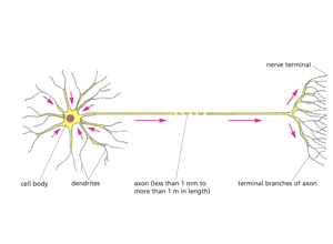
RIV-ULTRA S04: Neuron structure at the compartment level. Use it to fix vocabulary before you meet anything ambiguous: for each compartment, recall from §1 which organelles you would expect present and, more usefully, which would be absent. Ribosomes ruling out axon is the highest-value entry in that table.
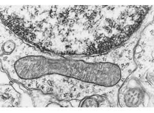
RIV-ULTRA S08: A somatic region — nuclear envelope with heterochromatin above, one long mitochondrion below. Use it to anchor the soma end of the compartment table in §1: rough ER and a nucleus put you in a cell body, and that is the one place where the ribosome cue is unambiguous rather than a judgment call.
RIV-ULTRA S09: The synapse cue set. Hold anything you would call to all three criteria in §2: a vesicle cluster at the apposition itself, a cleft of uniform width, and a density on the receiving side. Dark contrast alone is the commonest beginner error — no vesicles, no synapse.
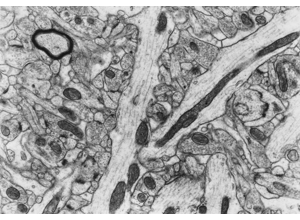
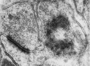
RIV-ULTRA S10: Vesicles and organelles at annotation scale. Check size against §1 before naming anything — clear synaptic vesicles run 35–50 nm and dense-core vesicles 80–120 nm, so this is a measurement rather than an impression. Remember that vesicle shape is partly a fixation artifact and is not independent of vesicle visibility.
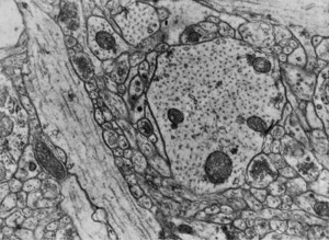
RIV-ULTRA S14: A comparative panel. Use it for the discipline §4 calls the whole game: pick two features that differ between profiles and ask whether they come from different cue families or share a failure mode. Two cues that degrade together under poor staining are one cue.
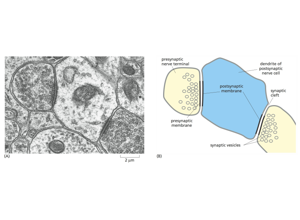
RIV-ULTRA S20: A two-panel reference — an EM micrograph with a 1 µm scale bar beside a labeled schematic naming presynaptic terminal, presynaptic and postsynaptic membranes, cleft, vesicles, and the postsynaptic dendrite. Read the schematic first, then find each labeled part in the micrograph beside it. That translation — idealized diagram to real noisy tissue — is the step §2's three criteria have to survive.
RIV-ULTRA S24: An advanced case for review. Build a full evidence chain in the form of the §4 worked example — geometry, organelle content, synaptic role, neighborhood — and stop at the point where the chain would need continuity across sections that a single still cannot supply.
Attribution: Pat Rivlin training materials (MICrONS proofreading deck).
5. Studio activity: ultrastructure consensus round (75 min)
Scenario. Your team is preparing a training-ready annotation subset for
segmentation QC. It deliberately contains borderline cases.
Independently label each patch: compartment, synapse status, confidence tier.
Record two supporting cues with their families and one uncertainty per patch.
Compare within the group; classify each disagreement as cue conflict, missing
context, or vocabulary mismatch.
Resolve what can be resolved; escalate genuine ambiguity with a written rationale.
Revise one rubric rule to reduce future disagreement of the type you saw most.
Outputs: consensus annotation sheet; disagreement log with counts by type; one
rubric revision with rationale.
Why step 5 matters. Vocabulary mismatch is usually the largest category on a first
run, and it is entirely fixable by better protocol wording. Teams that run this loop
two or three times typically see inter-annotator agreement rise substantially without
anyone becoming a better microscopist — the gain comes from the protocol, not the eye.
That is the scalability lesson of this unit.
Assessment rubric
Not yet
Proficient
Strong
Evidence quality
Single-cue calls presented as definitive
Two cues per call
Two cues from different families, with independence argued
Synapse criteria
Calls from contrast alone
Applies all three criteria
Applies all three plus persistence; correctly rejects adherens junctions and tangential membranes
Confidence
Missing or inconsistent
Tiers applied consistently
Tier justified against the operational definitions; uncertain rate is reasonable, neither zero nor excessive
Inference discipline
“Excitatory synapse”
“Putatively excitatory (asymmetric)”
Names the assumption and proposes corroboration via presynaptic cell type
Error analysis
Counts errors
Classifies disagreements by type
Converts the dominant disagreement type into a concrete protocol revision
Common errors and how to recover
Single-slice overconfidence. Recover: make scrolling a mandatory step in the
protocol, not a suggestion. Add a checkbox to the annotation sheet.
Stacking dependent cues. Recover: label each cue with its family. Three cues from
one family is one cue.
Forcing labels. Recover: define and reward the uncertain tier; track the uncertain
rate per annotator as a calibration statistic, not a performance penalty.
Label drift along a long trace. Recover: build in periodic re-checks — every N
micrometers of tracing, re-verify the compartment call from scratch rather than
carrying the earlier decision forward.
Reading artifact as biology. Recover: keep the Unit 03 artifact catalog open.
When something is anomalous, ask whether its shape follows tissue or follows the
section/tile/scan geometry.
The norm behind this unit
Some of what this unit teaches is technique. Some of it is professional norm — the
things experienced people do without being asked, and which nobody states out loud
because they assume you already know. Those are worth naming, because they are
distributed unequally by background rather
than by ability.
From this unit:
“Uncertain” is a real answer and should be rewarded out loud.
An annotator who never says “no” is not calibrated. Learners suppress uncertainty because they read it as failure; a facilitator has to say the opposite explicitly, once, early.
Two cues from different families, or it is not high confidence.
Three observations that share a failure mode are one observation. Nobody states this, and it is the single most common reasoning error in annotation.
The collected set, and why making these explicit is a fairness intervention rather than
etiquette, is in the hidden curriculum.
What this unit does not cover
Systematic axon-vs-dendrite classification, which gets its own treatment in Unit 06;
glial identification in Unit 07; and how these calls feed proofreading triage in
Unit 08.
Use at least two independent ultrastructure cues for each call.
Tag and escalate ambiguous regions with documented rationale.
Capability development brief
Capability target: Interpret ultrastructural features reproducibly to distinguish compartments and synaptic context.
Required expertise
Cellular neuroanatomist (organelle and compartment interpretation)
Senior proofreader (decision consistency under ambiguity)
Training lead (annotation rubric design)
Core concepts to teach
Compartment cues: Features such as mitochondria density, microtubules, vesicle pools, and membrane morphology.
Synaptic context: Interpreting cleft, vesicles, and postsynaptic density together rather than in isolation.
Confidence tagging: Marking uncertain calls to prioritize expert review.
Capability exercise
Ultrastructure Consensus Round - Build consistency in compartment and synapse labeling. The studio activity above is the graded version of this exercise; do that one.
Assessment artifacts
Compartment annotation rubric with confidence levels.
Inter-rater agreement report on a shared patch set.
Related concepts
Ultrastructure Annotation
Use compartment, organelle, and synaptic cues to make reproducible interpretation decisions.