Human cortex · H01
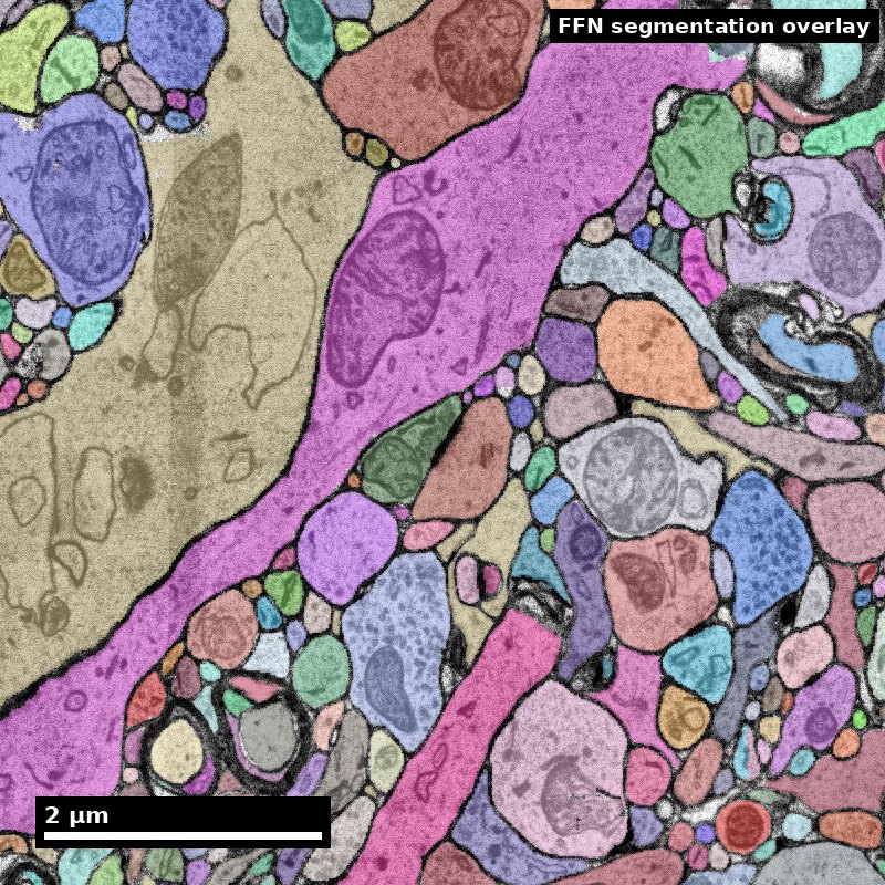
Object segmentation over electron microscopy
H01 release · Lichtman Lab / Harvard & Connectomics at Google · CC BY 4.0
Shapson-Coe et al. (2024) · doi:10.1126/science.adk4858
Technical Course · Unit 07
Glia
Astrocytes, oligodendrocytes and microglia in EM — and why a single glia–neuron merge is one of the most expensive errors in the volume.
Orientation: the stake
Glia occupy roughly 20–40% of cortical volume.
Source: Pat Rivlin training materials (MICrONS proofreading deck), RIV-GLIA S01.
Figure description and teaching notes
Set the stake before anyone starts looking: a glia–neuron merge does not merely add a branch — it drags a neuron's arbor past synapses it never contacted, so the false-input yield per micrometre of merged path is unusually high.
Each glial class has one near-diagnostic feature. Learn those three first: astrocyte — glycogen granules; oligodendrocyte — the darkest nucleus in the field; microglia — a dense elongated nucleus plus lysosomal content.
Astrocyte morphology in a synaptic neighbourhood
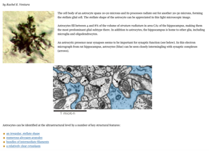
An astrocytic process is space-filling; a neurite is a tube.
Source: Ventura RE, "Astrocytes," SynapseWeb (Kristen M. Harris Lab, UT Austin), synapseweb.clm.utexas.edu/astrocytes. © The University of Texas at Austin; no reuse licence is stated — confirm permission before this deck is shared outside the course.
Figure description and teaching notes
Read cross-sectional shape before anything else. An astrocytic process takes whatever contour is left over after the neurites pack; a neurite holds a tube shape of its own. That cue survives weak staining better than glycogen granules do, which makes it the one to reach for where everything else fails.
Other astrocyte cues from unit §2: pale "watery" cytoplasm with few organelles; glycogen granules 20–30 nm, very dark, clustered (neurons do not contain them); perisynaptic processes under 100 nm thick; endfeet flattened against vessels; no vesicles, no PSDs, few microtubules.
Real data: an astrocytic process
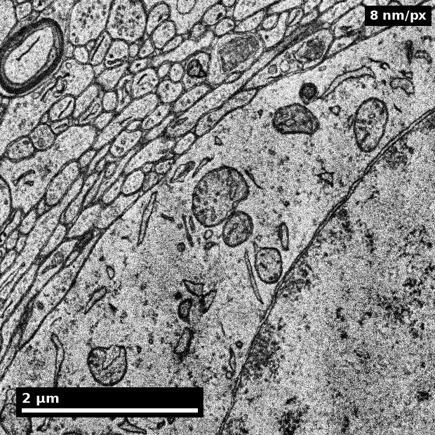
- Pale cytoplasm, irregular space-filling outline that wraps its neighbours.
- Labelled 'astrocyte' by H01's subcompartment model.
Source: H01 release (Lichtman Lab, Harvard; Connectomics at Google), CC BY 4.0 · Shapson-Coe et al. 2024, doi:10.1126/science.adk4858 · site render.
Figure description and teaching notes
Instructor script: "Trace the outline with your finger. Does it hold its own shape, or does it fill the gaps the neurites left?"
Use the unit's check-yourself: a pale, irregular profile between three neurites, no glycogen visible. Probably astrocytic — medium confidence at best. Absence of glycogen is weak evidence because granules are clustered and sparse. Better evidence, in order: space-filling behaviour over several sections; no vesicles or PSD on it; continuity to an endfoot; microtubules at higher magnification.
Real data: cortical vasculature
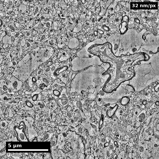
- H01 contains ~230 mm of blood vessels across ~1 mm³.
- Vessels are a common site of merge errors — glia wrap them closely.
Source: H01 release (Lichtman Lab, Harvard; Connectomics at Google), CC BY 4.0 · Shapson-Coe et al. 2024, doi:10.1126/science.adk4858 · site render.
Figure description and teaching notes
Walk the unit's worked example here: a pale profile a few hundred nanometres thick, pressed flat against a capillary, currently assigned to a nearby dendrite. Rule out synaptic participation first — even when the vascular context is shouting the answer. Glycogen absent: weak evidence, noted not spent. Over 5–10 sections the contour spreads to track the vessel wall: space-filling. Context: a flattened expansion covering a vessel is an endfoot. Dismiss microglia for cause — the parent process is pale and organelle-poor, not inclusion-rich.
Call: astrocytic endfoot, high confidence; the dendrite assignment is a glia–neuron merge and goes above conspicuous splits in the queue.
Microglia cues
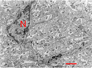
Weight nuclear evidence above process shape.
Source: Pat Rivlin training materials (MICrONS proofreading deck), RIV-GLIA S09.
Figure description and teaching notes
Look for the pairing that carries the call: a dark, often elongated or bean-shaped nucleus with heterochromatin clumped against the envelope, plus lysosomal and phagosomal content in dense cytoplasm with long, narrow ER cisternae. Microglia are the least stereotyped of the three classes because morphology tracks activation state — which is why nuclear evidence outranks process shape, and why a microglia call needs a nucleus or a lysosome-rich parent process, never a fragment.
Myelin-context interpretation
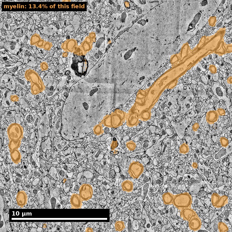
- Myelin is the fastest route to an oligodendrocyte identification: follow a sheath back to the soma that owns it.
- A merge across a myelinated axon costs more than one across an unmyelinated process: the axon it corrupts is long-range.
Source: H01 release (Lichtman Lab, Harvard; Connectomics at Google), CC BY 4.0 · Shapson-Coe et al. 2024, doi:10.1126/science.adk4858 · site render.
Figure description and teaching notes
The panel highlights every myelinated axon the H01 myelin mask finds in one grey-matter field — a small minority of profiles in cortex. Instructor cue: ask how myelin context changes proofreading priority. Context cues from the protocol: the inner or outer tongue of a myelin sheath, or a paranodal loop, is a high-confidence oligodendrocyte call.
The three classes, schematically

Source: Holly Fischer, Wikimedia Commons, File:Glial_Cell_Types.png. CC BY 3.0 Unported.